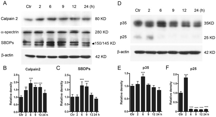
Figure 3. Protein levels of calpain 2, SBDPs, p35/p25 in cultured rat retinal neurons following GTs.
(A) Representative immunoblots showing the changes in calpain 2 and SBDP levels in cell extracts obtained from normal (Ctr) and glutamate-treated (0.5 mM for 2, 6, 9, 12 and 24 h) groups. (B, C) Bar charts summarizing the average densitometric quantification of immunoreactive bands of calpain 2 (B) and SBDPs (C) in Ctr and glutamate-treated groups, respectively. (D) Representative immunoblots showing the changes in p35 and p25 levels in cell extracts obtained from Ctr and glutamate-treated (0.5 mM for 2, 6, 9, 12 and 24 h) groups. Note that the immunoblots for p25 were over-exposed to make them clearer. (E, F) Bar charts summarizing the average densitometric quantification of immunoreactive bands of p35 (E) and p25 (F) in Ctr and glutamate-treated groups, respectively. Note that p25 expression was sharply increased following 2 h treatment, but decreased to a very low level with longer treatments. All data are normalized to Ctr. n = 6 for each group, ** p<0.01, *** p<0.001 vs Ctr, one-way ANOVA.

