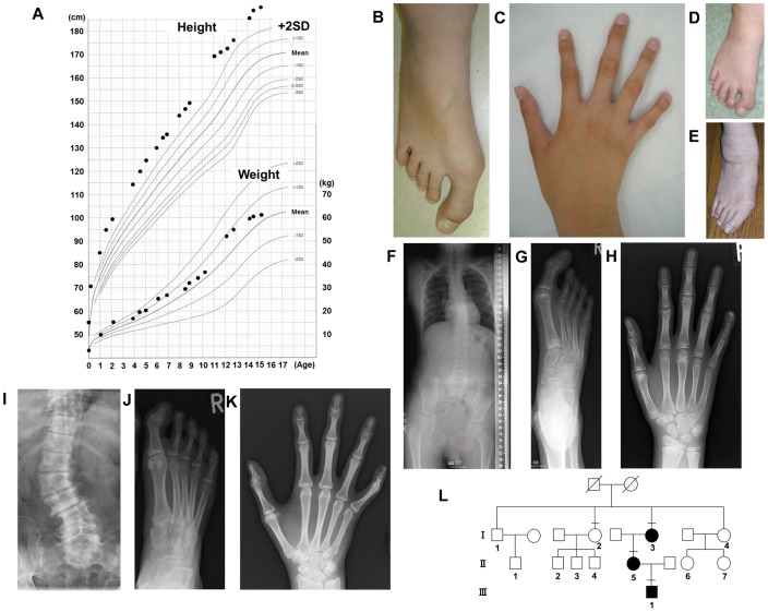
Figure 1. Clinical manifestations of the family.
(A) Growth curve of the proband. The X- and Y- axis indicate age (year) and height (cm) or weight (kg), respectively. The bold line represents the mean for Japanese males. (B, C) Photographs of the proband’s right foot (B) and hand (C). The great toe was markedly long and wide. The fifth distal phalanx of the hand also showed minor clinodactyly. He has given written informed consent to publication of his photographs. (D, E) The right foot of the mother (D) and grandmother (E), respectively. The mother’s great toes were surgically shortened at 15 years of age. The grandmother’s great toes were large in early life and shortened due to arthrogryposis with aging. They have given written informed consent to publication of their photographs. (F–H) Radiographs of the proband’s skeleton. His spine showed mild scoliosis (F). The great toe was markedly long and wide (G), and the fifth distal phalanx of the hand showed minor clinodactyly (H). (I–K) Radiographs of the mother. Severe scoliosis and lumbar vertebra fractures (I) as well as a markedly long and wide great toe (J), and minor clinodactyly in the fifth digit of the hand (K) were observed, like in the proband. (L) Family tree. III-1, II-5, and I-3 indicate the proband, mother, and maternal grandmother, respectively, who had the phenotype. I-2 indicates a maternal grand-aunt, who had no phenotype and was included in the present study.

