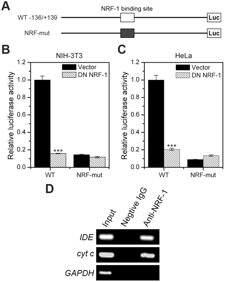
Figure 4. The NRF-1 binding motif in the mouse IDE promoter is functional.
(A) Representation of wild-type (−136/+139 WT) and the NRF-1 binding site-mutated (NRF-mut) luciferase reporter plasmids of the mouse IDE promoter. (B) and (C) Dominant negative NRF-1 represses IDE promoter activity. NIH-3T3 and HeLa cells were transiently co-transfected with wild-type (-136/+139 WT) or NRF-1 binding site-mutated (NRF-mut) IDE reporter plasmids (0.4 µg) and Renilla luciferase plasmid (4 ng) along with or without dominant negative (DN) NRF-1 expression plasmids (0.4 µg). Twenty-four hours after transfection, cells were lysed, and the luciferase activity was examined. Firefly luminescence signal was normalized based on the Renilla luminescence signal. (D) ChIP. NRF-1 binding to the IDE promoter in NIH-3T3 cells was determined by ChIP. The promoter of cytochrome c (cyt c) is used as a positive control for NRF-1 binding, while the promoter of GAPDH acts as a negative control.

