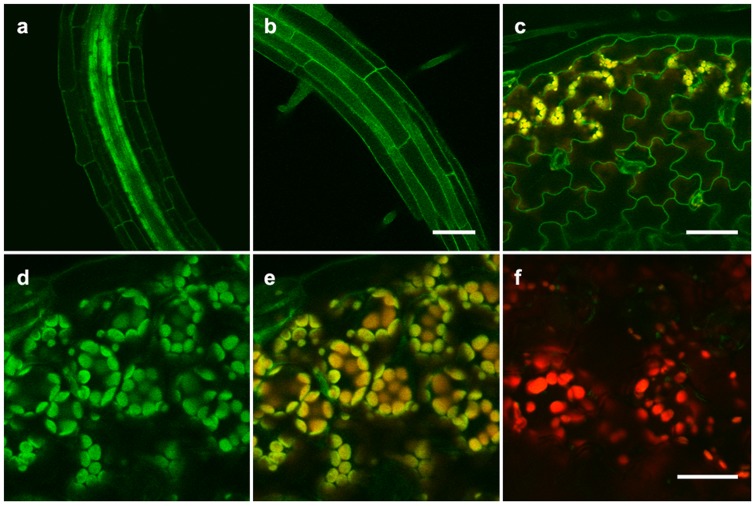
Figure 6. GFP-HvHMA2 localizes to the plasma membrane (PM) and chloroplasts of Arabidopsis.
(a) The PM is marked in all root cells within the root-hair initiation zone. Cells of the stele appear very bright at this stage. (b) In more mature root regions, the PM is very bright. Scale bar (a and b) = 50 µm. (c) In cotyledons, GFP-HvHMA2 marks the PM of epidermal cells (green) and the chloroplasts of mesophyll cells (orange). Orange colouring in chloroplasts results from overlay of the GFP and chlorophyll autofluorescence signals. Scale bar = 50 µm. (d–e) Higher magnification of chloroplast localization in cotyledons. GFP and chlorophyll autofluorescence are overlain in (e). (f) Chlorophyll autofluorescence (red) from cotyledon mesophyll cells of a untransformed control plant overlain with green channel emission collected as in (e). Note that very little green signal appears in wt chloroplasts. Scale bar (d–f) = 25 µm.

