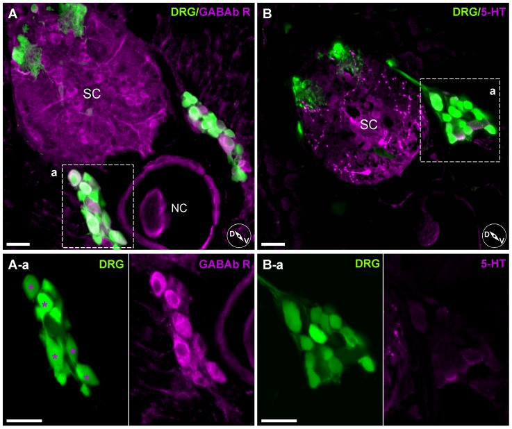
Figure 11. Heterogeneous expression pattern of anti-GABAb receptor antibody staining on DRG neurons.
A, Labeling with an anti-GABAb R1 antibody. Enlarged images (A-a, left and right) show only a specific subset of DRG neurons were labeled (asterisks). B, Labeling with an antibody against 5-HT. 5-HT antibody binding was mostly in spinal cord tracts rather than DRG neurons somata (enlarged box B-a). Inset cartoons represent orientation of images. D, dorsal; V, ventral; SC, Spinal Cord; NC, Notochord. Scale bars represent 20 µm.

