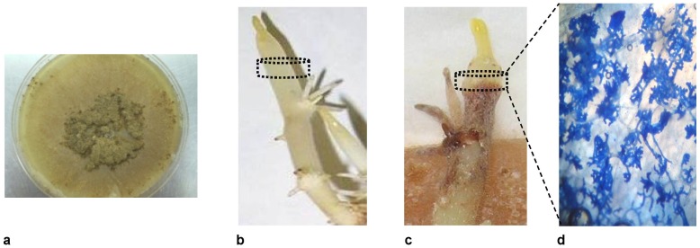
Figure 1. Rhizoctonia solani AG3 culture used as the inoculum (a), healthy and non-infected sprout (b), infected potato sprout 72 h after inoculation with the pathogen (c).
The portion of the sprout between the dashed lines was subjected to metabolomics analyses. In infected sprouts, samples included a small portion of the necrotic lesion. A necrotic region showing hyphae and infection cushions magnified at 600× (d).

