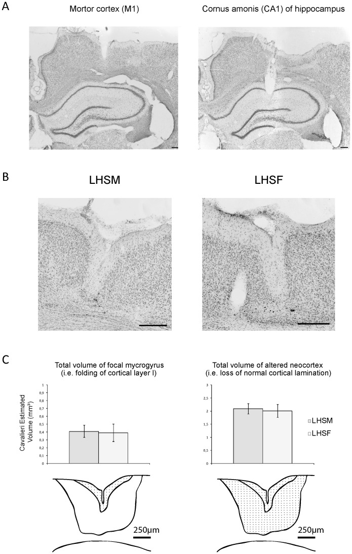
Figure 1. Photomicrographs of cresyl violet sections and Cavalieri's cortical lesion volumes estimations in the adult rats.
In A), a confirmation example of the bipolar electrodes placement for EEG recordings in a LHSF+T rat is shown in motor cortex M1 (left panel) and in the cornus ammonis region one (CA1) of the hippocampus (right panel). B) Cresyl violet stained coronal section through the dysplasic lesion in sensorimotor cortex of P120 rat brains, male (left) and female (right), that received a freeze-induced lesion at P1 and hyperthermic seizure at P10 (LHS). Photomicrographs are showing a similar and well-formed, four-layered microgyrus in the middle of each panel for both genders. In C), (Top panel) histograms of the volume estimations in the LHSM versus LHSF groups using the Cavalieri's principle for the focal mycrogyrus (left) and total amount of altered neocortex (right). (Bottom panel) diagrams illustrating the region of interest for sampling in each case highlighted with grid points. All Scale bars = 250 µm.

