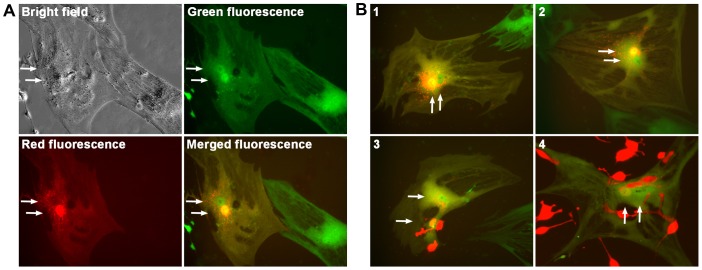
Figure 3. Confirming cancer-stromal cell fusion.
Representative cancer-stromal cell fusion events from the co-culture of RL-1 cells with hMSC-GFP cells are shown. A, A single fusion event at day 7 is shown in bright field, green fluorescence, and red fluorescence. The green and red fluorescence images are merged (merged fluorescence) to show the two nuclei of different fluorescence. B, Merged fluorescence images for 4 additional fusion events are shown, with events 1 and 2 recorded at day 7, and 3 and 4 at day 14. Arrows are used to indicate nuclei. All the images are shown at 200× magnification.

