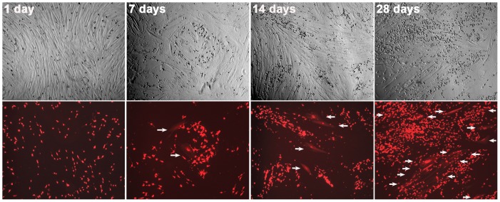
Figure 4. Time-dependence of cancer-stromal cell fusion.
Co-cultures of RL-1 and HPS-15 cells were observed weekly for frequency of cell fusion. For each view, a phase contrast image (top) and red fluorescence image (bottom) are shown. Arrows are used to indicate cancer-stromal cell fusion events. All the images are shown at 40× magnification.

