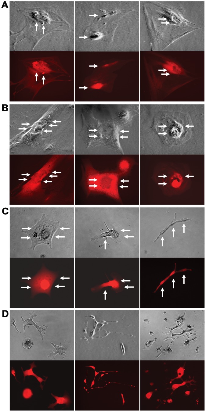
Figure 5. Tracking the fate of cancer-stromal hybrids.
Representative morphologies of the hybrid cells during colony formation are shown. A, Two weeks into the culture, most of the hybrids contained two nuclei of similar fluorescence. No cell division was observed. B, Four weeks into the co-culture, hybrid cells adopted atypical morphology with multiple nuclei. No cell division was observed. C, Six weeks into the culture, the remaining hybrid cells became thin or narrow, with multiple nuclei in segments of the cell. D, Eight weeks into the culture, cell division became prevalent. The cell division was abnormal because it produced daughter cells in varied shapes and with reduced viability. For each view, a phase contrast image (top) and red fluorescence image (bottom) are shown. When necessary, arrows are used to indicate nuclei. All the images are shown at 200× magnification.

