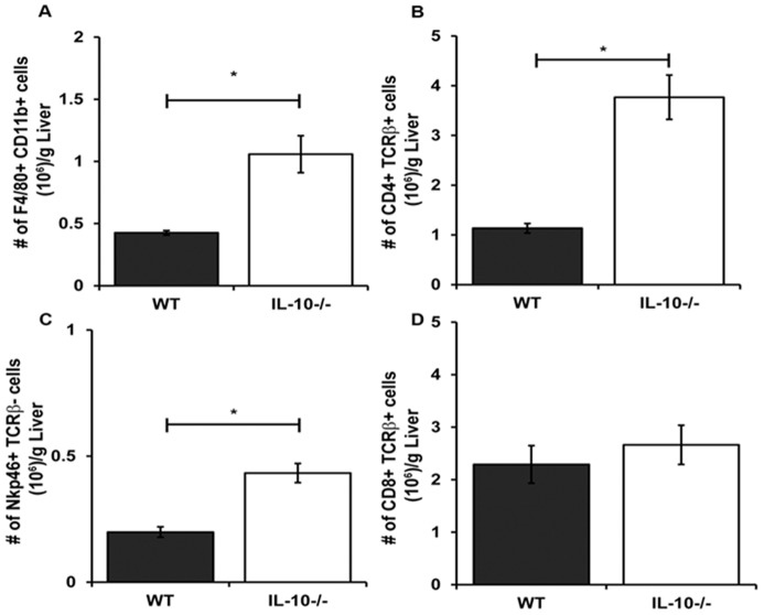
Figure 5. Persistence of effector cells in MCMV infected livers in the absence of IL-10.
The numbers of (A) macrophages, (B) CD4+ T cells, (C) NK cells, and (D) CD8+ T cells in the livers of WT and IL-10−/− mice infected for 9 days were determined by flow cytometry. The mean numbers of cells per gram liver ± SE are the combined data of four independent experiments (n = 12–13 mice per group). Asterisks denote statistically significant differences between WT and IL-10−/− groups, where p values are ≤0.05 (Student's T test).

