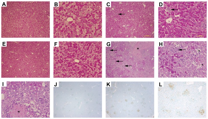
Figure 7. Liver pathology during acute MCMV infection in the absence of IL-10.
Livers from (A, B, C, D) C57BL/6J (WT) and (E, F, G, H) IL-10−/− mice that were (A, B, E, F) uninfected or (C, D, G, H, I, J, K, L) infected with MCMV for 5 days were harvested, paraffin embedded, and sectioned for PAS staining. Inflammatory foci (arrows, C, D, G & H), necrosis (asterisks, G and I), councilman bodies (black arrowheads, H), are marked. TUNEL positive cell clusters are shown in (J) Day 5 WT and (K and L) IL-10−/− livers. A, C, E, G, J & K were visualized at ×4 magnification. B, D, F, H, & I at ×20 magnification. L was visualized at ×10 magnification.

