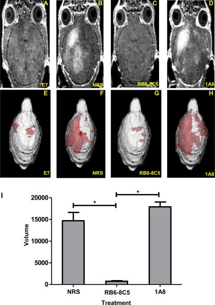
Figure 5. Neutrophil depletion with mAb 1A8 does not preserve vascular integrity as measured by 3D volumetric analysis of gadolinium enhancement visible on T1 weighted MRI.
Gadolinium enhanced T1 weighted MRI images showing the extent of vascular permeability in a representative (A) 7-day TMEV-infected mock E7 peptide administered C57BL/6 mouse not undergoing PIFS. In B–D, C57BL/6 mice undergoing PIFS were treated with (B) normal rat serum, (C) RB6-8C5, or (D) 1A8. E–H show 3D transparency rendering of gadolinium enhancing areas generated in Analyze 10.0 in the same animals. Red areas represent subvolumes with gadolinium enhancement. The intensity of red areas is influenced by the overall thickness of underlying gadolinium enhancing volume and by distance from surface. (I) Quantification of the 3D volume of vascular permeability in each of the 3 treatment groups. Treatment with anti-GR-1 mAb RB6-8C5 (n=5) significantly reduced vascular permeability as seen through decreased volumes of gadolinium enhancement when compared to treatment with normal rat serum (n=6) (p<0.05) and treatment with anti-Ly-6G mAb 1A8 (n=3) (p<0.05). Significance between groups was determined by an ANOVA followed by Dunn's method of multiple comparison. Error bars indicate SEM.

