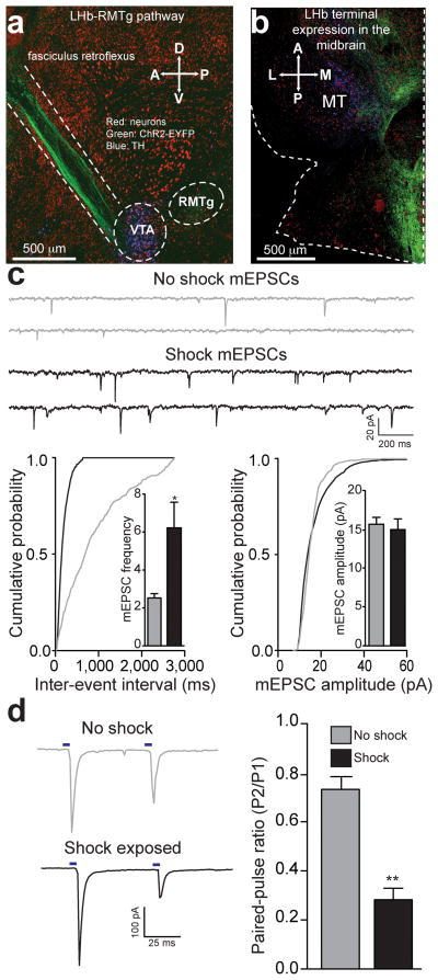
Figure 1. Acute unpredictable foot shock exposure enhances LHb-to-RMTg glutamate release.
(a) Sagittal confocal image showing expression of ChR2-EYFP (green) in the LHb-to-midbrain pathway via the fasciculus retroflexus fiber bundle following injection of the viral construct into the LHb. Midbrain TH+ dopamine neurons are shown in blue. A, Anterior; P, Posterior; D, Dorsal, V, Ventral. (b) Horizontal confocal image showing the distribution of LHb terminals in the midbrain. M, medial; L, lateral. (c) Top: representative mEPSC traces recorded from neurons from mice immediately following either 0 or 19 unpredictable foot shocks. Bottom Left: Representative cumulative mEPSC inter-event interval probability plot. Inset: Average mEPSC frequency was significantly increased in neurons from shock exposed mice (t(13) = 2.88, p = 0.01). Bottom Right: Representative cumulative mEPSC amplitude probability plot. Inset: Average mEPSC amplitude was not altered in RMTg neurons from shock exposed mice (t(13) = 0.12, p = 0.91). (d) Left: Representative optically evoked paired-pulse ratios from LHb efferents onto RMTg neurons. Right: Average paired-pulse ratios showing that paired-pulse ratios at LHb-to-RMTg synapses were significantly depressed from mice that received foot shocks (t(14) = 3.56, p = 0.003). n = 8 cells/group. All error bars for all figures correspond to the s.e.m. * indicates p < 0.05 and ** indicates p < 0.01 for all figures.

