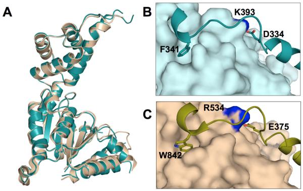
Figure 1.
Comparison of the two existing spastin structures. A) Superposition of human spastin (deep teal) and Drosophila spastin (wheat) AAA domains. (B) The loop between a1 and a2 (shown as ribbon representation) is held in the open configuration by a number of interactions with the ATPase domain (shown as surface), including the hydrophobic and ionic interactions seen above. (C) Similar interactions are seen in the nucleotide-free Drosophila spastin structure.

