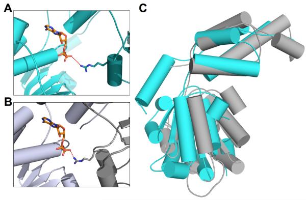
Figure 3.
Comparison of spastin monomer with hexameric AAA+ protein, FtsH. (A) Spastin (cyan) nucleotide binding pocket with modeled ADP molecule from FtsH. Arginine finger side chain shown as sticks and distance between arginine amine and beta phosphate of ADP shown as dashed line. (B) Interaction between the arginine finger and nucleotide of FtsH (C) Superposition of human spastin (cyan) and FtsH (grey) four-helix bundle domains, highlighting the difference in relative orientation of the HBD and NBDs of these structures.

