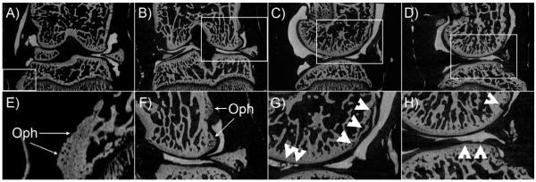
FIGURE 5.
Contrast enhanced μCT images of tibiofemoral joints after surgical destabilization of medial meniscus. A and B) coronal plane displaying the medial meniscus out of place. C and D) sagittal views of the DMM model showing ulceration and erosion of tibial and femoral cartilage respectively. E and F) magnified views of A and B showing the formation of osteophytes in the tibia and femur. G and H) disruption and ulceration of femoral and tibial cartilage.

