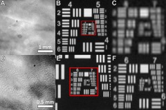
Fig. 2.
Microscopic methylene blue stained ex vivo mouse colon and USAF bar target images viewed at (a) and (b) and (d) and (e). Mucosal crypt structure was resolved between the two sets of images, indicating a desired surface magnifying chromoendoscopy modality imaging resolution between . Parts (c) and (f) show enlarged views of boxed regions in parts (b) and (e), respectively.

