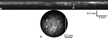
Fig. 8.
Optical coherence tomography (OCT) image scanning the full 30 mm of the in vivo mouse colon (a), and surface magnifying chromoendoscopy (SMC) image taken at one location in the mouse colon (b). The layered structures of the colon, including mucosa (M), submucosa (SM), and muscularis propria (MP) are easily identified in the OCT image, while arrows in the SMC image point to individual colonic crypts, and the white dotted line corresponds to the scan line of the specific OCT image.

