Fig. 3.
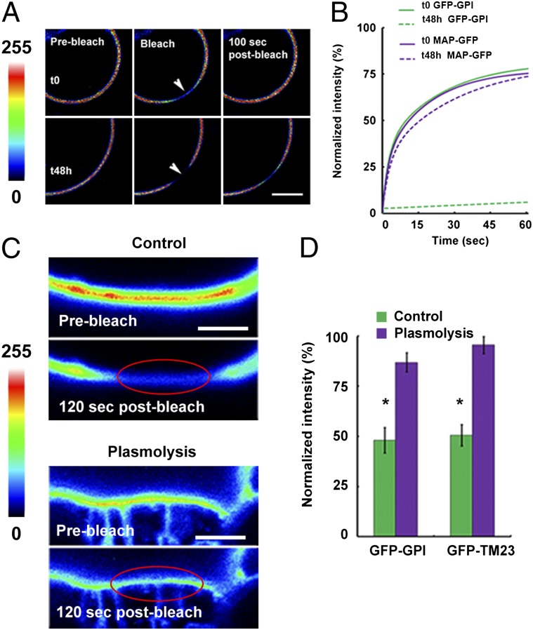
The cell wall limits lateral mobility of plant PM proteins. (A) Protoplasts expressing GFP-GPI. Fluorescence recovers quickly in freshly prepared protoplasts, but the protein is much less mobile once the cell wall has regrown for 48 h. Arrowheads indicate the bleached region. Color scale indicates pixel intensity from 0 (black) to 255 (brightest intensity possible). (Scale bar: 5 μm.) (B) Mobile fraction of GFP-GPI and MAP-GFP in freshly prepared protoplasts (t0) and after cell-wall regeneration (t48h). The mobile fraction (I60s) of GFP-GPI decreased significantly as the cell wall was neosynthesized (I60s GFP-GPI t0 = 79.9 ± 3.2%; t48h = 2.6 ± 0.6%; P < 0.001, t test), but that of MAP-GFP did not change. (C) GFP-GPI is relatively immobile in FRAP experiments in control cells but becomes mobile in cells plasmolyzed with 0.5 M mannitol. Color scale indicates pixel intensity from 0 (black) to 255 (brightest intensity possible). Red ellipse marks the bleached region. (Scale bars: 2 μm.) (D) Plasmolysis induces a highly significant increase in fluorescence recovery (*P < 0.001 for both I60s GFP-GPI control vs. plasmolysis and I60s GFP-TM23 control vs. plasmolysis; t test).

