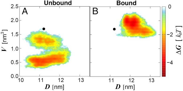
Fig. 3.
Shift of LSD1/CoREST conformational sampling in solution upon H3-histone-tail binding. Data from the 0.5-μs (A) unbound and (B) H3-bound ensembles is summarized in terms of the SWIRM–SAINT2 interdomain distance, D, and the H3-pocket volume, V. Color-coding ranges from light blue (less favorable free energy) to red (most favorable free energy). Sampling starts from the X-ray reference structure (black circle). D and V values from the X-ray reference structure are representative of all X-ray models deposited in the Protein Data Bank to date.

