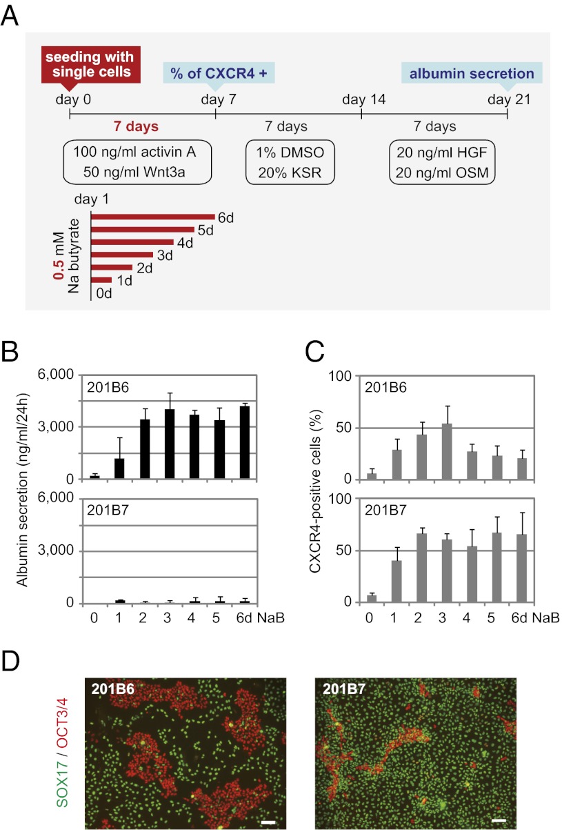
Fig. 3.
Close comparison of the propensity for hepatic differentiation between sibling hiPSC lines 201B6 and 201B7. (A) Schematic representation of the modified protocol used for hepatic differentiation. In this protocol, hiPS/ESCs were enzymatically digested into single cells and plated on Matrigel-coated dishes. For endodermal cell induction, the cells were cultivated with activin A and Wnt3a for 7 d. A total of 0.5 mM sodium butyrate (NaB) was supplemented from day 1 for various durations (0–6 d). (B) Albumin secretion level after 21 d of hepatic differentiation. The error bars indicate the SD (n = 3). (C) Percentage of CXCR4-positive cells after 7 d of endodermal differentiation as determined by flow cytometry. Error bars indicate the SD (n = 3). (D) Immunostaining of SOX17 and OCT3/4 after 7 d of endodermal differentiation. NaB was added for 3 d. (Scale bar: 100 μm.)

