Fig. 2.
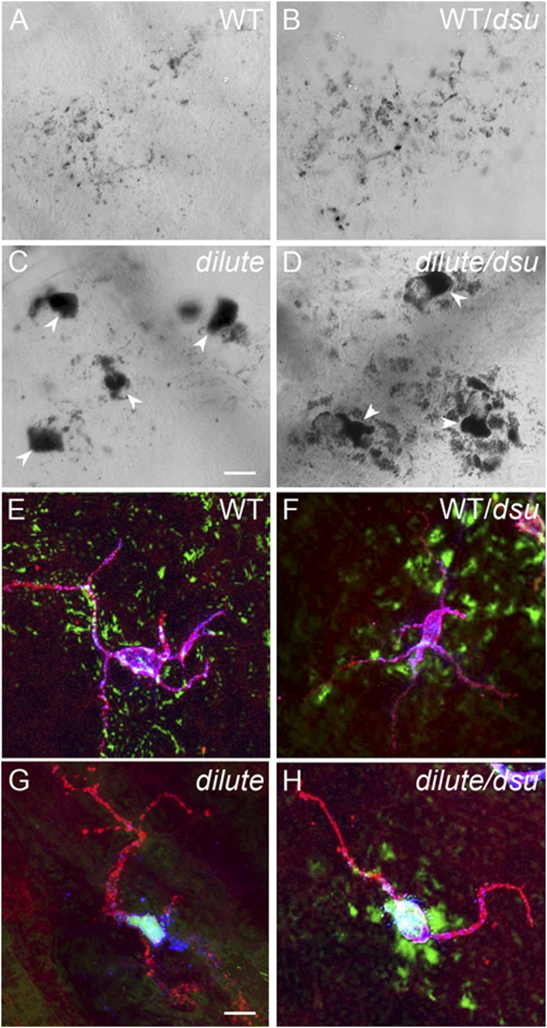
Melanosomes escape readily from the center of dilute/dsu ear skin melanocytes but not from the center of dilute ear skin melanocytes. Shown are bright-field images of ear skin from WT (A), WT/dsu (B), dilute (C), and dilute/dsu (D) mice. Also shown are images of ear skin from WT (E), WT/dsu (F), dilute (G), and dilute/dsu (H) mice that had been stained with an antibody to the plasma membrane receptor for Kit to define the shape of melanocytes and with an antibody to the melanosomal membrane protein TRP1. The anti-Kit signal is in red, black pigment is pseudo colored green, and the anti-TRP1 signal is in blue (yielding a blue-white color inside melanocytes because of the overlay with the red and green signals). (Scale bars: 10 μm in A–D; 7 μm in E–H.)

