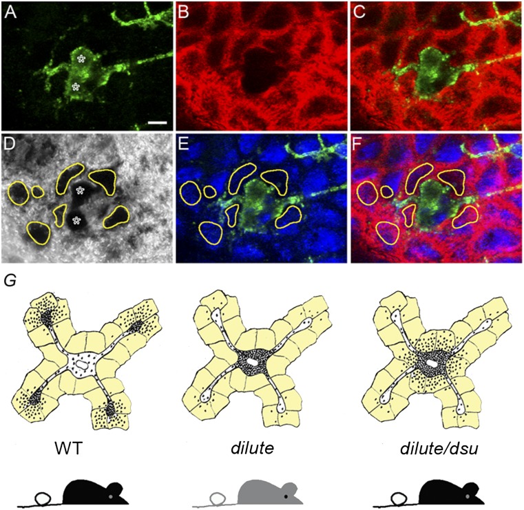
Fig. 3.
Pigment that escapes the center of dilute/dsu melanocytes is inside adjacent keratinocytes. Shown are images of ear skin from a dilute/dsu mouse that had been stained for Kit to reveal the shape of melanocytes (green signal in A, C, E, and F), keratin 14 to reveal the presence of keratin filaments within keratinocytes (red signal in B, C, and E), and DAPI to reveal the positions of nuclei (blue signal in E and F) and the distribution of pigment (black in the transmitted-light image in D). (A–C) Two adjacent melanocyte cell bodies in green (asterisks in A) surrounded by keratinocytes, which appear as red ovals because of the keratin filaments present in their peripheral cytoplasm (D) In the transmitted-light image, the dramatic accumulations of pigment that surround the two melanocyte cell bodies (marked with asterisks) are circled in yellow. (E) Overlay of this transmitted-light image (including the yellow circles) with the DAPI and Kit signals. (F) Overlay of this transmitted-light image (including the yellow circles) with the DAPI, cKit, and keratin 14 signals. (Scale bar: 7 μm.) (G) Cartoon depicting the basic concept supported by the images in Figs. 2 and 3, i.e., that dsu rescues the coat color of dilute mice without rescuing the defect in intracellular melanosome distribution exhibited by dilute melanocytes by allowing the melanosomes accumulated in their central cytoplasm to be transferred readily to the keratinocytes that immediately surround the melanocyte’s cell body (the keratinocytes in the cartoon are colored light yellow).

