
Fig. 7.
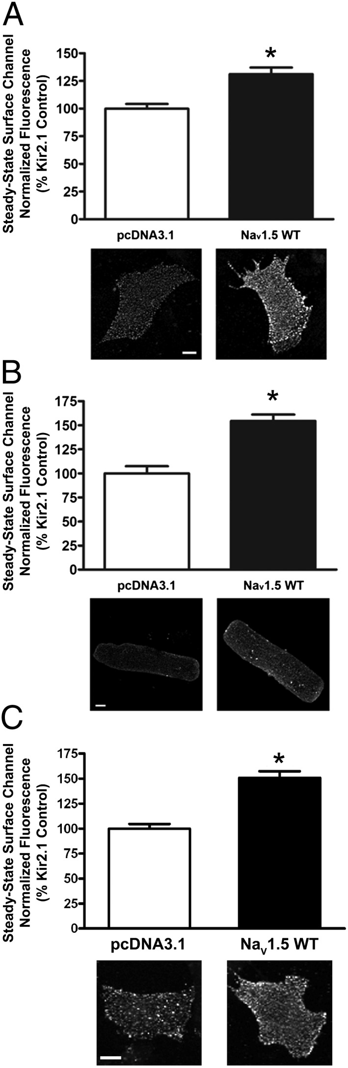
Coexpression of Kir2.1-pHluorin with NaV1.5 promotes cell surface expression of Kir2.1-pHluorin. Quantification of steady-state cell surface Kir2.1-pHluorin in cells cotransfected/coinfected with either empty pcDNA3.1 and DsRed vectors or plasmids/adenoviruses encoding NaV1.5 and DsRed. Cells were live cell labeled with anti-GFP antibody before incubation for 20 min at 37 °C. Cell surface channels were then labeled as described in SI Materials and Methods. Representative images of surface Kir2.1-pHluorin channels for each condition and cell type are shown. (Scale bars: 10 μm.) *P < 0.005 as determined by unpaired Student t test. (A) Rat neonatal ventricular myocytes (n > 58 myocytes). (B) ARVMs (n > 75 cells). (C) HL-1 cells (n > 56 cells). DNA plasmid transfection was used for rat neonatal ventricular myocytes and HL-1 cells; adenoviral-mediated overexpression was used for ARVMs.