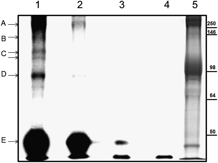
Fig. 4.
Labeling of ATP pool-associated membrane proteins with [α-32P]-N3-ATP. This was accomplished following the same protocol used for N3-ATP. Labeled proteins were separated by SDS/PAGE and analyzed with a Cyclone phosphoimager. Lanes 1–4 show phosphoimages of ghost proteins after separation. Lane 5 is the Coomassie blue-stained counterpart. The proteins tentatively identified are indicated with arrows. Possible protein candidates for the labeled bands are: A, β-spectrin and/or ankyrin; B, Ca2+ pump (Mr ∼140 kDa) or fragments of spectrin/ankyrin; C, α-subunit of the Na+/K+ pump (Mr ∼110 kDa); D, band 3; and E, actin.

