Fig. 1.
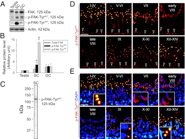
Expression of phosphorylated FAK and the stage-specific expression pattern of p-FAK-Tyr407 in adult rat testes. (A) Immunoblots show the expression of FAK and its phosphorylated forms in adult rat testes, Sertoli cells (SC; isolated from 20-d-old rat testes and cultured for 5 d), and germ cells (GC; freshly isolated from adult rat testes and used within ∼2 h). Actin served as a protein loading control. (B) Histogram summarizes immunoblotting results as in A from several independent experiments. Each data point was normalized against the corresponding actin level, and the protein level in the testis was arbitrarily set as 1, against which statistical comparison was performed. Each bar represents a mean ± SD of n = 3–4. *P < 0.05; **P < 0.01 vs. testis; 1-way ANOVA followed by Dunnett’s test. (C) Immunoblot shows the specificity of the anti–p-FAK-Tyr407 antibody (Table S1). (D) Immunofluorescence staining of p-FAK-Tyr407 in frozen sections of adult rat testes (red) is shown. (E) Merged images with nuclei stained with DAPI (blue) are shown. Each image shows a cross-section of the seminiferous epithelium from a tubule with its stage annotated with roman numerals I through XIV. (Insets) Boxed areas in selected images in E were magnified. p-FAK-Tyr407 was localized in the basal compartment near the basement membrane, consistent with its localization at the BTB (open arrowheads in D) at all stages. It was also highly expressed, most predominantly at the concave side of elongated spermatid heads in stage VII to early stage VIII tubules (Insets in E), corresponding to structures known as apical TBC at the Sertoli cell-elongating spermatid interface, which is the “degenerating” apical ES (2), but the signal diminished rapidly to a nondetectable level in late stage VIII. In addition, p-FAK-Tyr407 appeared in vesicle-like structures near the tubule lumen except at stage VIII, apparently as part of the cytoplasmic droplets that were shredded during spermiogenesis, perhaps representing “degraded” p-FAK-Tyr407. (Scale bar: D and E, 40 μm.)

