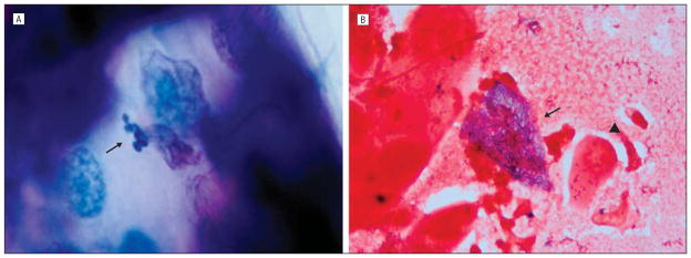
Figure 2.
Microscopic examination of a diagnostic epithelial scraping. A, Case 1. There are numerous 2.0×1.0–μm organisms in the cytoplasm of an epithelial cell (arrow) (Giemsa, original magnification ×250). B, Case 2. An epithelial cell is distended with 2.0×1.0–μm organisms in its cytoplasm (arrow) compared with a normal cell (arrowhead) (Gram, original magnification ×250).

