
Figure 3.
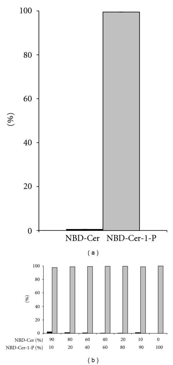
Separation of NBD-C6-Cer and NBD-C6-Cer-1-P via NH2-SPE. A mixture containing NBD-C6-Cer and NBD-C6-Cer-1-P, both at 5 μM (a) or at a total concentration of 5 μM but with a variable ratio (b) in the assay medium was separated via NH2-SPE, as described in Section 2. The eluted fractions were dried, resolubilized in chloroform/methanol (1/2, v/v) and the lipids were separated on silica G TLC plates (chloroform/acetone/methanol/acetic acid/water, 10/4/3/2/1, v/v), followed by fluorescence scanning of the spots (NBD-C6-Cer (black bars); NBD-C6-Cer-1-P (grey bars).) The result is expressed as percentage of total fluorescence in the elution fraction (a) mean ± SEM; n = 5; (b) single experiment).