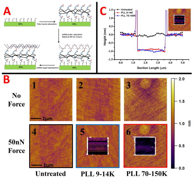
Figure 5.
A schematic of the surface functionalization of the HfO2 surface for microRNA (DNA analogue) sensing is shown in A. AFM images of the HfO2 and poly-l-lysine layers of different molecular weights are shown in B. Tapping mode images with no force applied (upper) for the different layers, and after a 50nN scratching force (bottom) are displayed. The scale bar for all AFM images is on the right. A cross section for the images with 50nN force applied is in part C. The cross sections are color coded to images in B with an inset representing the cross sectional area.

