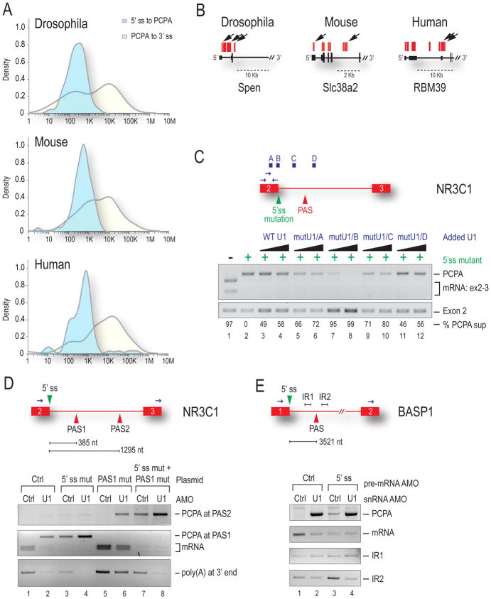
Figure 4. U1 bound outside the 5’ss is required to suppress PASs >1kb away.
(A) Distribution of the log10 distance at which PCPA occurred was measured in each organism from the start of the poly(A) tail back to the upstream 5’ss (light blue) or downstream to the 3’ss (yellow). See also Supplemental Figures S2–S4 and Table S2. (B) Arrows indicate multiple PCPA sites per gene. (C) HeLa cells were transfected with WT NR3C1 mini-gene (lane 1) or one containing a 5’ss mutation (lanes 2–12) and increasing amounts of mutant U1 that base-pairs to one of four locations along the pre-mRNA (above in blue). PCPA in the intron was detected by 3′RACE with RT-PCR of exon 2 serving as the loading control. Percent suppression (PCPA to exon 2 ratio) was normalized to lane 2. (D) Control or U1 AMO were transfected along with WT NR3C1 mini-gene (lanes 1 & 2) or NR3C1 in which the PAS1 [385 nt] sequence in the intron was duplicated (PAS2) and placed 1295 nt downstream of the 5’ss. Mini-genes in which the 5’ss (lanes 3 & 4), PAS1 (lanes 5 & 6) or both (lanes 7 & 8) were mutated are indicated. 3′RACE was performed as in (C). (E) Control, U1 or an AMO to the 5’ss of the endogenous BASP1 gene were transfected as indicated. PCPA at the PAS 3.5 kb downstream of exon 1 was measured by 3′ RACE. RT-PCR on intronic sequences upstream (IR1) and downstream (IR2) of the PAS is shown.

