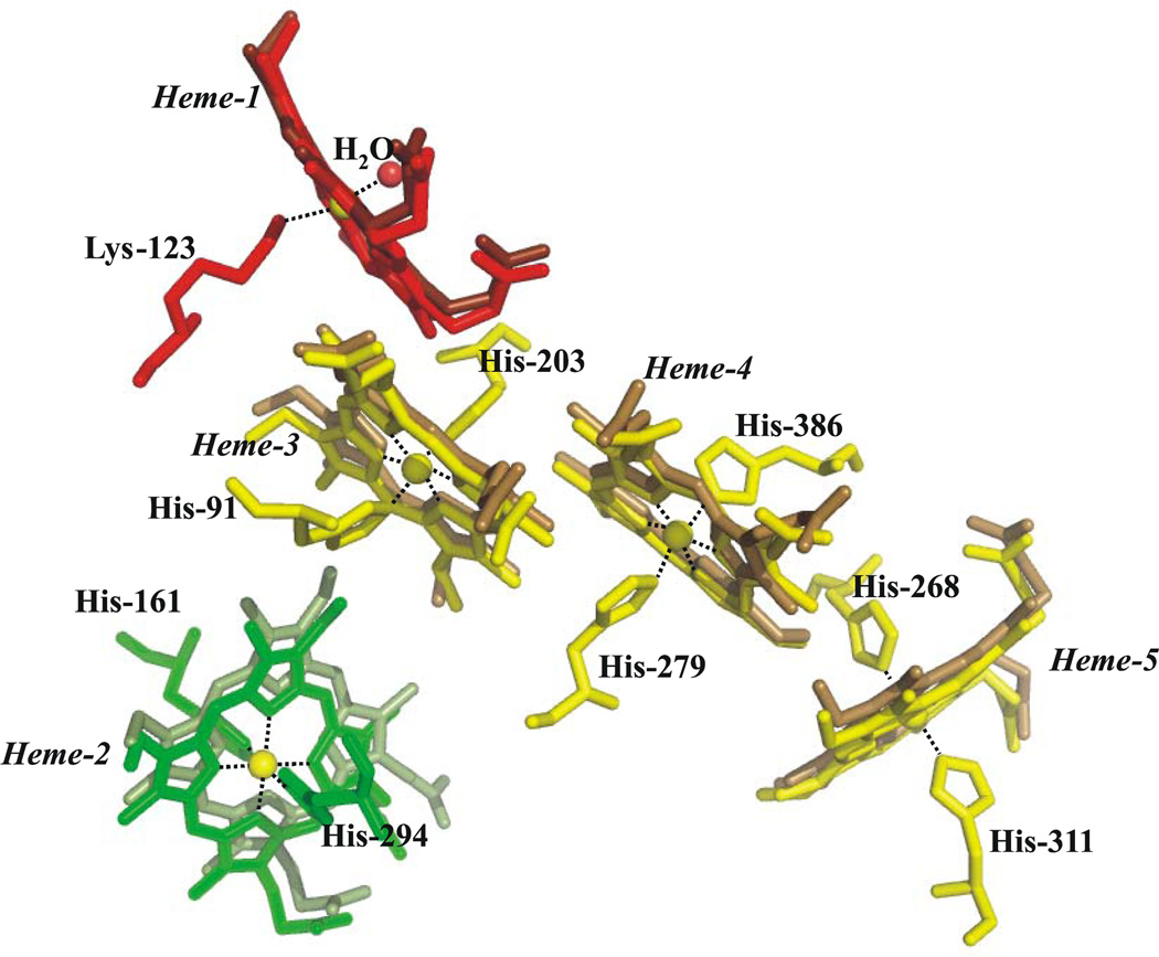
Figure 6.
Comparison between the heme arrangement within a monomer of S. oneidensis ccNiR (lighter shade) and that within a monomer of E. coli (darker shade). Subunit A is shown. Irons are shown in yellow, while the heme color scheme matches that of Fig. 1.

