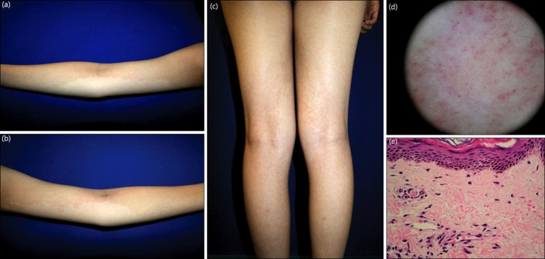
Fig. 1.
(a~c) Brownish hyperpigmented patches on both arms and legs in case 1. (d) Dermoscopy showing multiple purpuric globules over an orange-brown background. (e) Histopathological analysis showing perivascular lymphocytic infiltrates with extravasation of erythrocytes in the papillary dermis (H&E, ×400).

