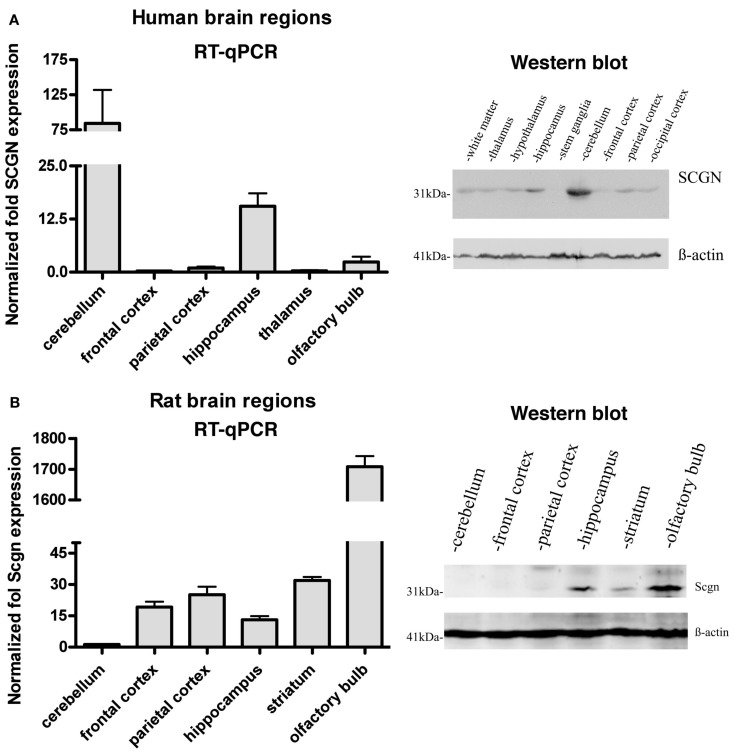
Figure 1.
Expression of Secretagogin in different regions of human and rat brain. Tissues from different brain areas were analyzed for relative Secretagogin gene and protein expression by RT-qPCR and Western blotting. (A) Human post-mortem tissues from three individuals were analyzed with regard to the following specific brain regions: cerebellum, frontal cortex, parietal cortex, hippocampus, thalamus, and olfactory bulb. Western blot analysis was performed from tissue of white matter, thalamus, hypothalamus, hippocampus, stem ganglia, cerebellum, frontal cortex, parietal cortex, and occipital cortex. Equal protein loading was verified by staining with β-actin antibody. (B) Rat brain tissues from three adult rats were dissected and analyzed from cerebellum, frontal cortex, parietal cortex, hippocampus, striatum, and olfactory bulb. A representative Western blot of equal amounts of protein from cerebellum, frontal cortex, parietal cortex, hippocampus, striatum, and olfactory bulb is shown on the right. β-actin immunostaining was again used as loading control.

