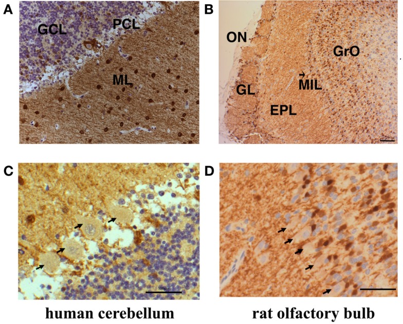
Figure 3.
Subcellular distribution of Secretagogin-positive cells in human cerebellum and rat olfactory bulb. Paraffin sections from human cerebellum (A) and rat olfactory bulb (B) were processed for Secretagogin immunoreactivity (DAB staining in brown) and counterstained with Mayer’s Hemalaun (blue). Human cerebellum reveals positive Secretagogin staining of interneurons in the molecular layer (ML) and the granule cell layer (GCL). Granule cells are immuno-negative and Purkinje cells bodies revealed faint positive staining (PCL). Actual expression of Secretagogin in Purkinje cells would need verification by single cell analysis. The rat olfactory bulb shows Secretagogin-positive cells in the glomerular layer (GL), the external plexiform layer (EPL), and the granular cell layer (GrO). The mitral cell bodies (indicated by an arrow, MIL) seem to be very weakly positive. Scale bars: 200 μm. Magnifications of the human Purkinje cell layer and rat mitral cells are shown in (C,D) respectively. Principal cells are indicated by arrows. Scale bars: 20 μm.

