Figure 4.
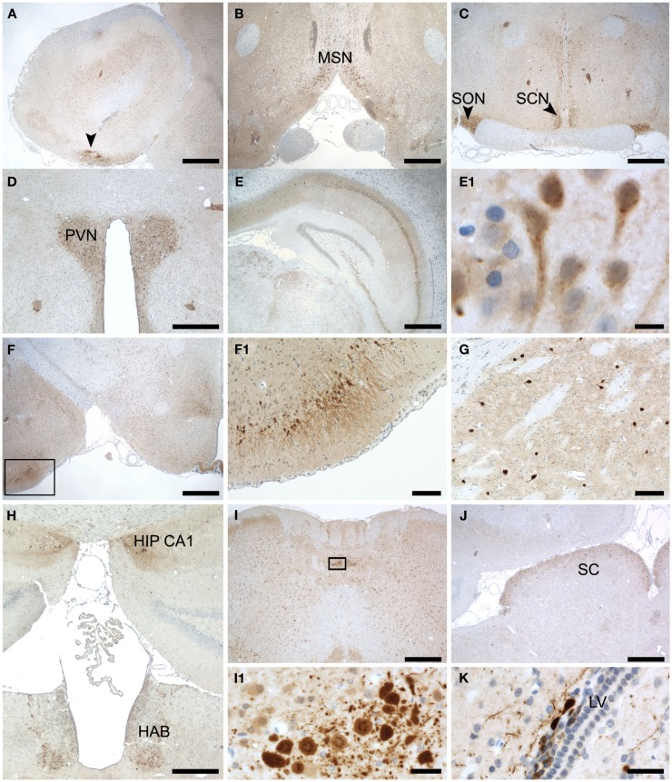
Subregional expression of Secretagogin in rat brain. Representative sections from rat brain were stained for Secretagogin immunoreactivity and counterstained with hematoxylin. Panels represent the following brain areas: (A) olfactory bulb, arrow pointing to a patch of strong Scgn+ cells. Scale bar: 500 μm. (B) Medial septal nucleus. Scale bar: 500 μm. (C) Supraoptic nucleus (SON) and Suprachiasmatic nucleus (SCN). Scale bar: 500 μm. (D) Paraventricular nucleus (PVN). Scale bar: 400 μm. (E) Hippocampus. Scale bar: 500 μm. (E1) Magnification of hippocampal neurons in CA1. Scale bar: 10 μm. (F) Lateral amygdaloid nucleus (indicated by box). Scale bar: 500 μm. (F1) Magnification of the region indicated in (F). Scale bar: 100 μm. (G) Cells in caudate putamen. Scale bar: 100 μm. (H) Panel reveals two Scgn+ areas: beginning of hippocampal CA1 region (HIP CA1) and habenula (HAB). Scale bar: 400 μm. (I) Medulla oblongata. Scale bar: 500 μm. (I1) Magnification of a strongly Scgn+ nucleus in medulla oblongata as indicated in (I). Scale bar: 25 μm. (J) Superior colliculus (SC). Scale bar: 500 μm. (K) Cells around the lateral ventricle. Scale bar: 25 μm.

