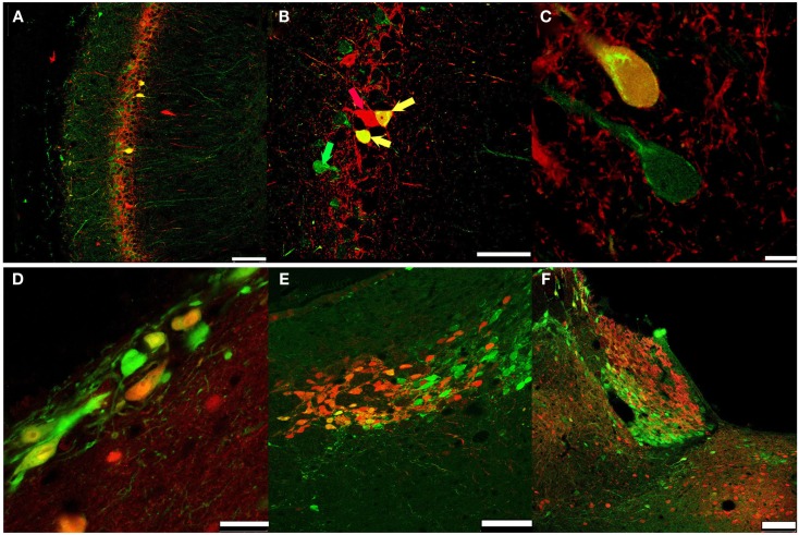
Figure 5.
Secretagogin and other CBPs. PFA-fixed rat brain slices were immunostained for Secretagogin (Scgn: green) and other CBPs (red) and processed for immunofluorescence. (A-C) Hippocampal CA1 region: Secretagogin (green), Parvalbumin (PV, red). (A) Scale bar: 100 μm. (B) Arrows indicate co-expression of Scgn and PV; green arrow: Scgn only, red arrow: PV only; yellow arrow: co-expression of Scgn and PV in a single neuron. Scale bar: 50 μm. (C) Larger magnification of Scgn/PV co-expressing neurons indicates a distinct subcellular distribution. Scale bar: 7.5 μm. (D) Secretagogin-positive neurons near the ventricle. Secretagogin (green), Calbindin D28k (red). Scale bar: 25 μm. (E) Habenula: Secretagogin (green), Calbindin D28k (red). Scale bar: 75 μm. (F) Habenula: Secretagogin (green), Calretinin (red). Scale bar: 100 μm.

