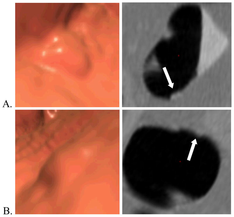
Figure 3.

Example of a hyperplastic polyp (arrows). Three-dimensional endoluminal (left) and two-dimensional cross-sectional (perpendicular to centerline) (right) (A) prone and (B) supine CT colonography images of a 8 mm flat hyperplastic polyp (white arrows) in the transverse colon of a 58-year-old man. The polyp’s widths and heights computed by automated software are (A) 6.4 and 3.5 mm and (B) 8.3 and 1.9 mm. For this polyp, there was a substantial change in both width and height between the supine and prone scans suggesting pliability.
