Figure 1.
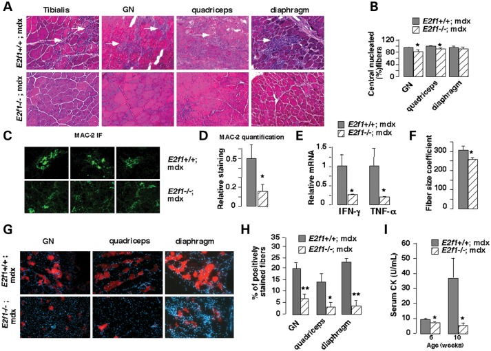
Decreased fiber damage in E2f1 −/−;mdx male mice. (A) Hematoxylin staining of different muscles [gastrocnemius (GN)] from E2f1+/+;mdx and E2f1−/−;mdx mice. Necrotic lesions (white arrowhead) are observed only in E2f1+/+;mdx mice. Representative micrographs of one out of four animals are shown. (B) The percentage of centrally located fibers was measured using hematoxylin staining of different muscle sections from three E2f1+/+;mdx and E2f1−/−;mdx mice. All values represent means ± standard error of mean (SEM). *P < 0.05; **P < 0.01 here and in subsequent figures. (C) Inflammatory infiltration in E2f1+/+;mdx and E2f1−/−;mdx mice, as demonstrated by Mac-2 immunofluorescence. Nuclei are stained with Hoechst. (D) Quantification of Mac-2 infiltration using Image J software. Intensity of the staining was corrected by total fiber area. (E) Quantitative mRNA expression of inflammatory cytokines in muscles of E2f1−/−; compared with E2f1+/+;mdx mice. (F) Cross-sectional area of individual fiber was measured on hematoxylin-stained sections of GN muscle and the coefficient of fiber size variability was calculated by averaging the standard deviation of data from three mice for each genotype. (G) Evans blue was injected i.p., and GN and quadriceps tissue sections were analyzed by fluorescence microscopy. (H) Quantification of Evan blue positive-stained fibers in the different E2f1+/+;mdx and E2f1−/−;mdx muscles as indicated. (I) Serum CK in E2f1+/+;mdx and E2f1−/−;mdx mice aged at 6 and 10 weeks.

