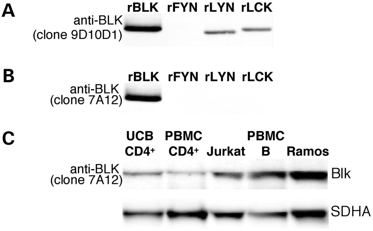
Figure 3.
Western blot analysis of Blk antibody specificity. Recombinant Blk (lane 1) and Src kinases Fyn, Lyn and Lck (lanes 2, 3 and 4, respectively) were run on an SDS–PAGE gel. Western blot analysis was performed using anti-Blk monoclonal antibody clones 9D10D1 (A) and 7A12 (B). (A) Anti-Blk clone 9D10D1 clearly detects rBlk, rLyn and rLck and is thus not specific for Blk. (B) The anti-Blk antibody clone 7A12 recognizes rBlk and does not label rFyn, rLyn or rLck. (C) Whole cell lysates were analyzed by western blot: lanes; 1, UCB CD4+ T cells; 2, adult peripheral blood CD4+ T cells; 3, Jurkat cells; 4, adult peripheral blood total B cells separated using MACS; 5, Ramos cells. Blk was detected using anti-Blk clone 7A12. The lower panel shows SDHA as a control for the amount of protein loaded.

