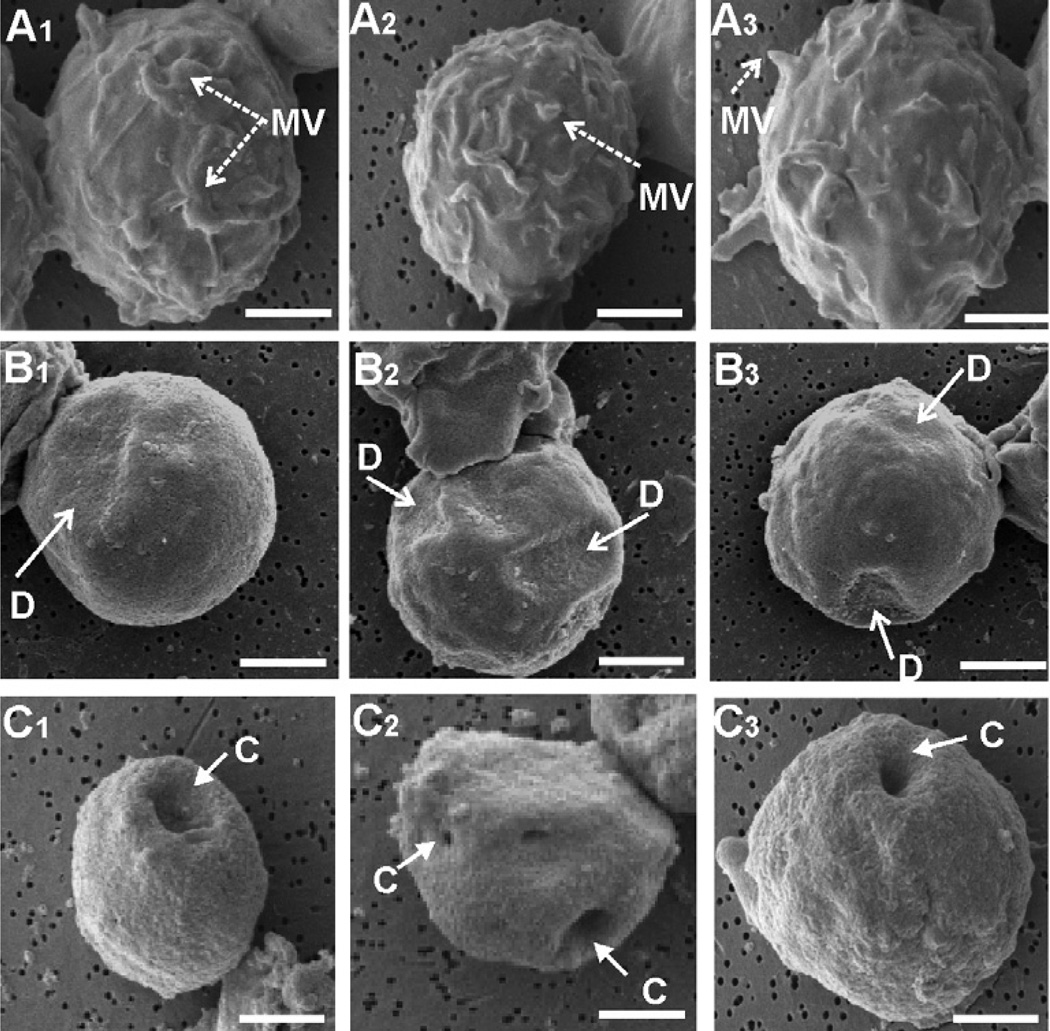
Fig. 2.
LtxA mediates collapse of microvilli on Jn.9 target cells. The samples were prepared for conventional scanning electron microscopy of an untreated control (Fig. 2A1–3) and experimental groups after 30 min (Fig. 2B1–3) and 3 h (Fig. 2C1–3) of incubation with LtxA (1 × 10⁻⁹ M). Images are representative of the unique phenotypes observed within each group in three independent experiments. All images were acquired at × 6000 and bars equal 2.5 µm. MV, microvilli; D, depression; C, cavity.

