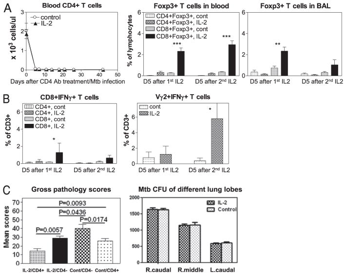
FIGURE 7.
Depletion of IL-2–expanded CD4+Foxp3+ Treg and CD4+ T effector cells reduced, but did not eliminate, IL-2–induced homeostatic protection against TB lesions. (A) A complete in vivo deletion of CD4+ T cells (left graph) by anti-CD4 Ab treatment during M. tuberculosis infection, and blockage of IL-2 expansion of CD4+Foxp3+ Treg (middle and right graphs). Data were collected on day 5 (D5) after the first (1st) and the second (2nd) IL-2 treatment as day 5 after each IL-2 treatment is the peak time for Foxp3+ T cell expansion (see Fig. 1). (B) CD4 depletion blocked the ability of IL-2 to expand CD4+ T effectors during M. tuberculosis infection. Effector CD8+ T and γδ T cells were detectable. (C) Left graph, CD4 depletion significantly reduced IL-2–induced resistance to TB lesions (see the first and second bars [from left] for comparison between IL-2–treated CD4-depleted [IL-2/CD4−] group and IL-2–treated CD4-not-depleted [IL-2/CD4+] group), but IL-2 treatments still led to detectable resistance to severe TB lesions in the absence of CD4+ T cells (see the second and third bars for comparison between saline-treated [Cont/CD4−] and IL-2–treated [IL-2/CD4−] groups). Another control group, saline-treated CD4-not-depleted (Cont/CD4+) group, was also included for comparison. (C) Right graph, No statistical difference in mean M. tuberculosis CFU counts from lung lobe homogenates between IL-2–treated CD4-depleted group (IL-2) and control saline-treated CD4-depleted group (Control). Note that extrathoracic TB lesions were seen in the control group, but not in the IL-2–treated group after CD4 depletion. Both IL-2 and control groups of macaques (8 animals) were treated with humanized OKT4A-depleting mAb at a dose of 50 mg/kg on days –5, 10, and 25 after M. tuberculosis infection. Intermittent IL-2 treatments, M. tuberculosis infection, and evaluations of gross pathology and bacterial burdens were essentially the same as those described above for those macaques without CD4 depletion. The data in this figure are compared between the groups based on our previous observation that control isotype Ab treatment of macaques during M. tuberculosis infection does not cause significant depletion or expansion of CD4+ T effector cells, CD4+Foxp3+ T cells, CD8+ T effector cells, or γδ T effector cells when compared with saline-treated macaques (Ref. 22 and data not shown). *p < 0.05, **p < 0.01, ***p < 0.001.

