
Figure 3. Susceptibility-weighted images of patient 6, 7, and 9.
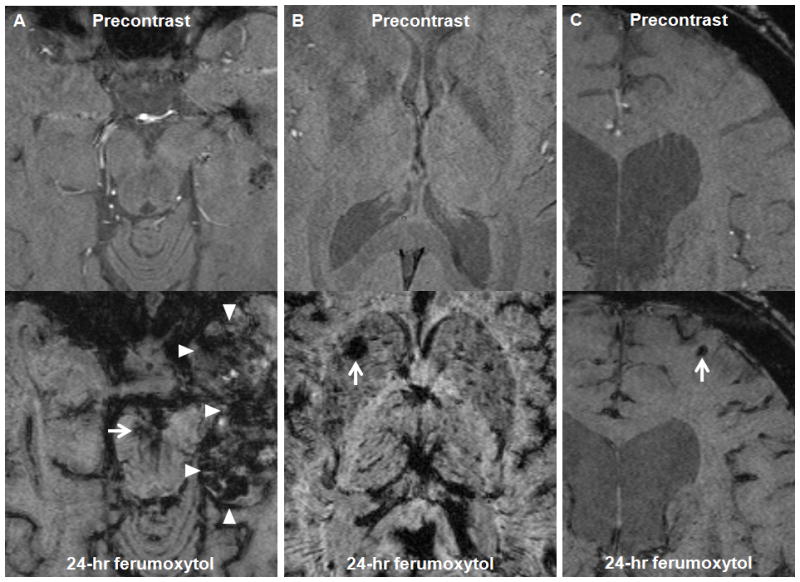
A–C, Axial susceptibility-weighted images of patient 6 (A), 7 (B), and 9 (C) obtained before and 24 hours after ferumoxytol administration. A, The ferumoxytol MRI shows a small capillary telangiectasia in the mesencephalon (arrow), while the precontrast image does not. Areas of decreased signal intensity are observed within the left temporal lobe tumor (arrowheads) 24 hours after ferumoxytol administration. B, The ferumoxytol MRI shows a capillary telangiectasia in the right basal ganglia (arrow), while the precontrast image does not. C, The ferumoxytol MRI shows a small cavernoma in the left anterior frontal lobe (arrow), while the precontrast image does not.