
Figure 4. Developmental venous anomaly.
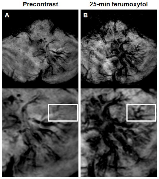
A–B, Axial susceptibility-weighted images obtained before (A) and 25 minutes after ferumoxytol administration (B). Albeit the left cerebellar lesion is visible on both the pre- and post-ferumoxytol susceptibility-weighted images, but the ferumoxytol scan demonstrates additional tributary veins (rectangle) compared with the precontrast sequence.