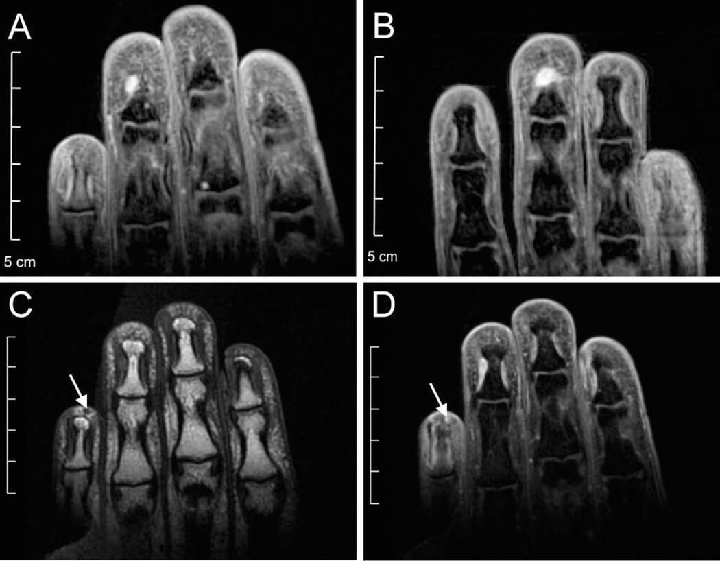
Figure 2.
MRI images from NIH–1. 35 year-old Hispanic woman with NF1 and severe pain in the third, fourth and fifth digits of both hands, exacerbated by cold temperatures, on disability. She also presented with signs and symptoms of the complex regional pain syndrome: allodynia, swelling and vasomotor changes. Over three years, she required multiple surgeries to treat recurrent and metachronous glomus tumors (Table S1). Prior to her first surgery, coronal T2-weighted MRI revealed a ~5 mm lesion in left F4 (A) and ~6 mm lesion in right F3 (B). Coronal T1-weighted pre-(C) and post- (D) gadolinium imaging revealed a ~1.8 mm lesion in left F5. All three lesions were glomus tumors on pathologic examination. No enhancing lesions were visible in other symptomatic fingers.

