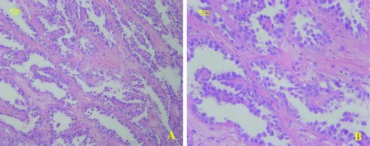
Figure 1.
Histological features of CDC in a representative section. (A) Typical irregular angulated tubular architecture associated with striking stromal desmoplasia. (B) High-powered view of CDC showing cancer cells with large pleomorphic nuclei and eosinophilic cytoplasm. Pathological tissue section No. 15397. Scale bars, 20 μm.

