
Fig. 2.
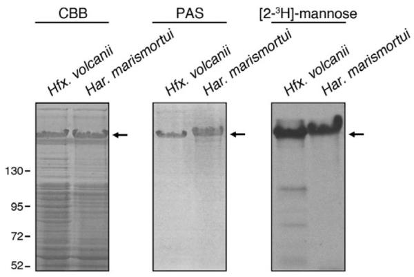
Like its Hfx. volcanii counterpart, the Har. marismortui S-layer protein is glycosylated. The protein contents of Hfx. volcanii (left) and Har. marismortui (right) cells grown to identical OD550 values separated by 7.5% SDS-PAGE and either Coomassieor PAS-stained [left (CBB) and middle panels respectively]. In addition, the protein contents of Hfx. volcanii (left) and Har. marismortui (right) cells challenged with [2-3H]-mannose were separated by 7.5% SDS-PAGE and processed for fluorography (right panel). In each panel, the Hfx. volcanii and Har. marismortui S-layer glycoproteins are indicated by an arrow. Molecular mass markers are shown on the left of the figure.