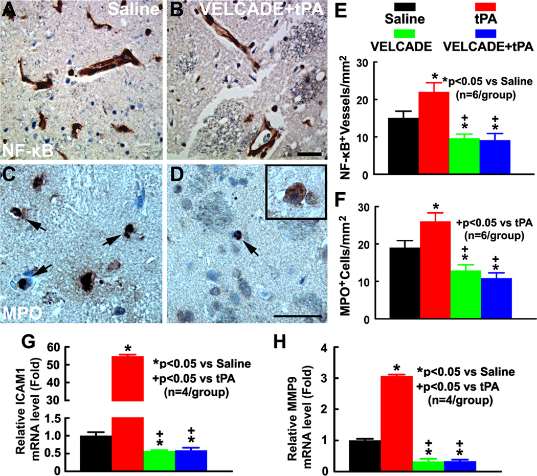
Figure 4. Treatment effects on proinflammatory gene expression and neutrophil accumulation.
Panels A and B show p65 (NF-κB) immunoreactive cerebral vessels from representative rats treated with saline (A) and the combination of VELCADE and tPA (B). Panels C and D show MPO immunoreactive cells from representative rats treated with saline (C), and the combination of VELCADE and tPA (D). MPO-positive cells having the typical polymorphonucleated neutrophil morphology(arrows, C, D) which can be easily distinguished from macrophages by their large size and apparent cytoplasmic inclusions (insert in D). Quantitative data (E, F) show of the number of NF-κB immunoreactive vessels (E) and MPO immunoreactive cells (F) in ischemic brain of the rat treated with saline, tPA, VELCADE, and VELCADE with tPA. Quantitative RT-PCR data show mRNA levels of ICAM1 (G) and MMP9 (H) on cerebral endothelial cells isolated by LCM in the ischemic hemisphere of the rat treated with saline, tPA, VELCADE, and VELCADE with tPA. Bars=50µm.

