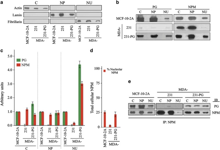
Figure 2.
Subcellular distribution of PG and NPM and their association in MDA-231 and MDA-231-PG cells. (a, b) MCF-10-2A (control), MDA-231 and -231-PG cells were grown in 150 mm culture dishes until confluent. Cell pellets (total of 108 cells) were processed for subcellular fractionation according to the Lamond's protocol to isolate the cytoplasmic (C), nucleoplasmic (NP) and nucleolar (NU) fractions. The purity of C, NP and NU fractions were verified by immunoblotting with actin, lamin and fibrillarin antibodies, respectively (a). Fractions were processed for immunoblotting with PG and NPM antibodies (b). The experiment was repeated five times with similar results and the data is representative of one such experiment. (c) Quantitation of the data presented in b. The amount of each protein in the cytoplasmic (C), nucleoplasmic (NP) and nucleolar (NU) fraction was normalized to the amounts of actin, lamin and fibrillarin, respectively, in the same sample extract relative to MCF-10-2A cells. PG and NPM values for MDA-231-PG transfectants are the average of multiple independent clones. (d) The ratio of the nucleolar/total cellular NPM detected in various cell lines. The value for MDA-231-PG transfectants is the average of multiple independent clones. (e) Cytoplasmic (C), nucleoplasmic (NP) and nucleolar (NU) fractions from MCF-10-2A (control), MDA-231 and -231-PG cells were prepared as described in Figure 2a and processed for immunoprecipitation using NPM antibodies. Immune complexes were resolved by SDS–PAGE and immunoblotted with antibodies to PG and NPM. Each experiment was repeated at least five times with similar results and the data is presented for one typical experiment.

