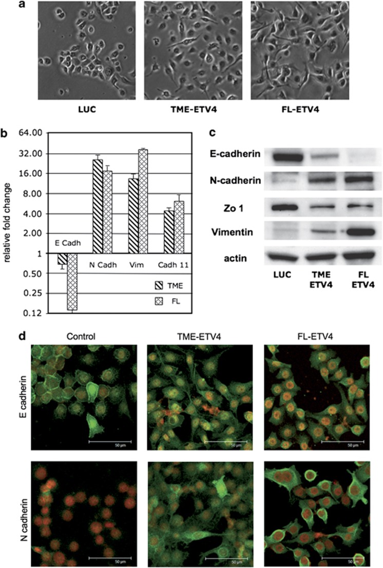
Figure 6.
ETV4 overexpression induces EMT in the RWPE. (a) Representative microphotographs of RWPE cells are shown. Almost all mock-transfected RWPE cells displayed a rounded epithelial cell shape with rare spindle-shaped cells. A large fraction of cells RWPE transfected with FL and TME vectors displayed a spindle-like shape. (b) Fold changes of expression levels (normalized to the housekeeping gene glyceraldehyde-3-phosphate dehydrogenase) of epithelial (E-cadherin) and mesenchymal (N-cadherin, vimentin and cadherin 11) markers in RWPE cells transfected with FL-ETV4 and TME-ETV4 plasmids compared with mock-transfected RWPE cells. Fold changes are plotted on a Log 2 scale. (c) Expression assessed by western blot analysis of epithelial (E-cadherin and zonula occludens) and mesenchymal (N-cadherin and vimentin) markers in RWPE cells transfected with mock (Luc), FL-ETV4 and TME-ETV4 plasmids. (d) Analysis by confocal microscopy of E-cadherin and of N-cadherin in RWPE cells transfected with mock, FL-ETV4 and TME-ETV4 plasmids. The luciferase-transfected RWPE cells do not express the mesenchymal marker protein N-cadherin and the epithelial marker protein E-cadherin is expressed and localized on the plasma membrane. In RWPE cells overexpressing ETV4, either TME or FL, the E-cadherin is reduced and has migrated from the plasma membrane to the cytoplasm; the N-cadherin is upregulated and it appeared in both the cytoplasm and plasma membrane.

